
Hands-On Dental Course | Level 2
The Pattern Behind the Breakdown
Occlusion, Function, and the Comprehensive Plan
Read the wear, the force, and the joint as one pattern, and the plan follows.
Level 1 established the records and the preliminary diagnosis. Level 2 completes the diagnosis by adding occlusion and function, and turns it into a plan. It teaches you to read occlusion as the functional system it is, because that is where most diagnoses stop short.
You will interpret wear facets by angle and pattern, determine how occlusal force is distributed and where it concentrates, and recognize the functional risk that predicts which restorations will fail and when. From that reading you will construct a treatment plan that addresses the cause rather than the presenting tooth, sequence it correctly, and explain to the patient why a single crown will not solve a problem the whole system produced. This level also introduces TMJ disorders and their classification, cephalometric and CBCT imaging, and the role of appliances, establishing the mechanism that the later levels build on.
Learning objectives
Upon completion, participants will be able to:
Analyze functional occlusion and identify the occlusal factors that determine diagnosis and prognosis.
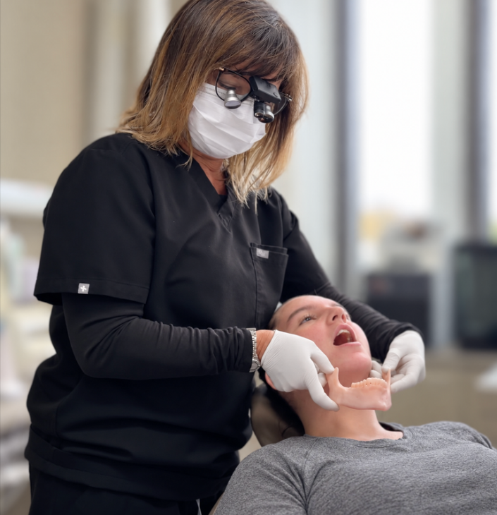
Assess mandibular position and take a bite registration that records it.
Interpret wear facets by angle, pattern, and location, and state what each indicates about function and force.
Evaluate how occlusal force is distributed across the arch and identify points of harmful concentration.
Apply force-management principles so that restorative work withstands the functional load placed on it.
Assess the TMJ and classify the findings: distinguish deviation from deflection and a click from a pop, and state what each indicates.
Describe the mechanism linking mandibular position, condylar seating, and disc position, and the role of an appliance within it.
Determine when a frontal or lateral cephalogram is indicated, and which view answers the clinical question.
Interpret craniocervical angulation, cervical curvature, hyoid position, and posterior airway on frontal and lateral cephalograms, and state how each acts on mandibular position and on the records taken.
Orient a CBCT volume and perform a basic reading, and recognize the findings that warrant further evaluation.
Synthesize the Level 1 records and examination with the occlusal findings into a complete diagnosis and a sequenced treatment plan.
Explain to a patient why isolated single-unit treatment is inadequate when multiple functional factors are present.
Course details
Location: Smiles by Bis
710 Dorval Dr #220, Oakville, ON L6K 3V7
Date: April 9 - 10, 2027
CE Credits: 14 (7.25 lecture, 6.75 participation)
Dentist tuition: $2,995 CAD
Hygienist or assistant: $980 CAD
Format: Two-day, in-person course
Class size: Limited to 16 participants
Included: Breakfast and lunch both days, Day 1 reception, course materials, and the Treatment-Planning Flowchart
Full refund less a $100 administrative fee more than 30 days before the course. 50% refund or one transfer 15 to 30 days before. No refund 14 days or less before; one transfer may be permitted.
CE approval: TMJ Whisperer Academy. Nationally Approved PACE Program Provider for FAGD/MAGD credit. Approval does not imply acceptance by any regulatory authority or AGD endorsement. 4/1/2026 to 3/31/2030. Provider ID# 434863.
Course content
-
Level 2 treats occlusion as a functional system, not a static contact pattern. You will learn to read occlusion in function: the contacts that guide the mandible, the interferences that redirect it, and the relationship between where the teeth meet and where the condyle seats. A diagnosis that omits the occlusion is incomplete, and a treatment plan built on an incomplete diagnosis moves in the wrong direction.
-
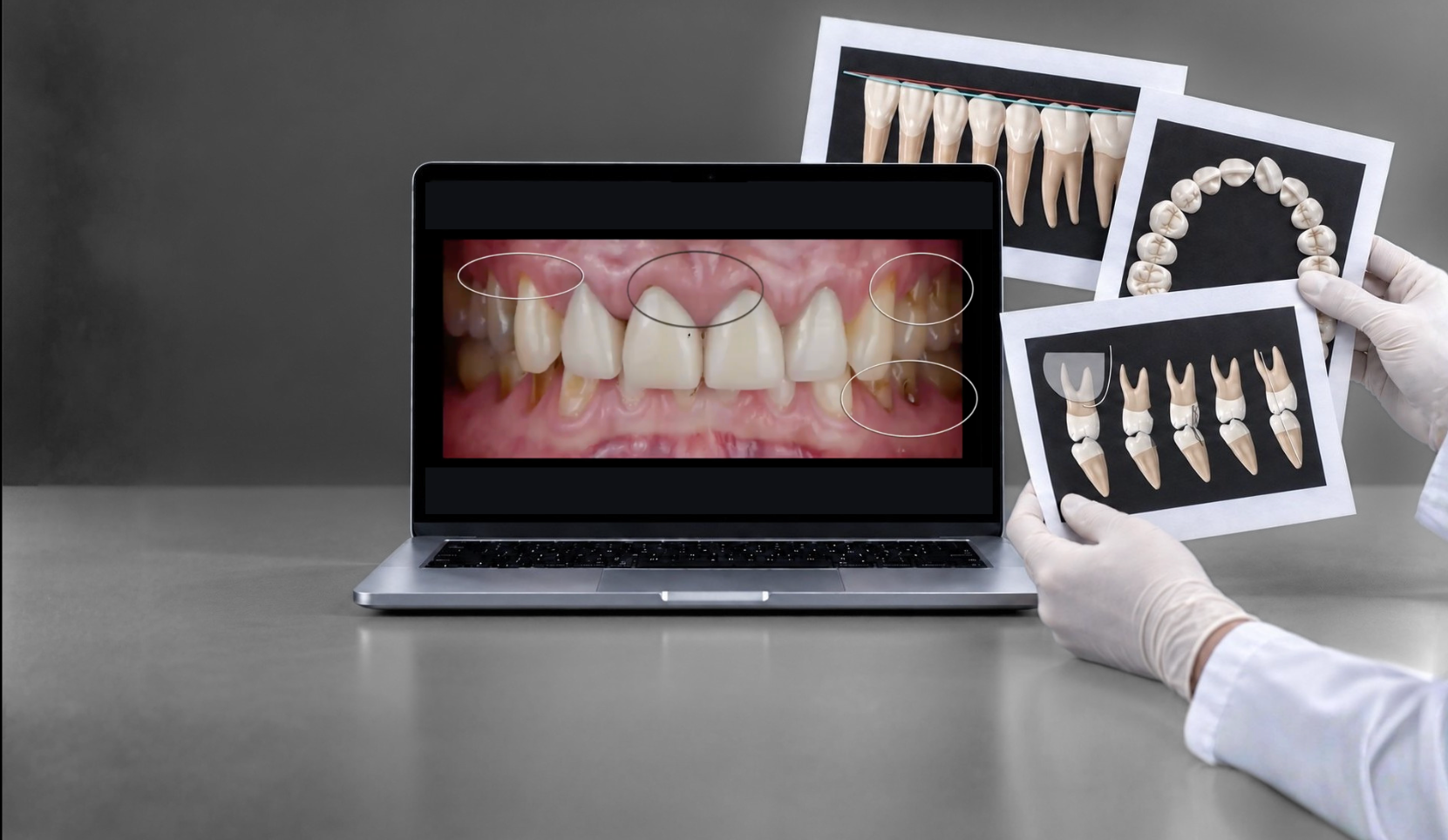
Wear facets are a physical record of how the mandible has moved and where force has been applied. Their angle, pattern, and location indicate the path of function and the sites of concentrated load. This module teaches you to read facets in detail: what a given angle indicates, what a pattern reveals about parafunction, and what both mean for the prognosis of any restoration placed into that environment.
-
Restorative work fails when it is loaded in ways it cannot withstand. This module teaches you to distribute occlusal force correctly across the arch, to identify where force concentrates, and to design treatment that manages that load so the dentistry lasts.
-
This module introduces the mechanism behind TMJ disorders. Mandibular retrusion leads to condylar distalization, which produces retrodiscal compression and, in turn, disc displacement. Understanding this chain is what allows you to connect a patient's occlusal findings to their joint symptoms rather than treating the two as separate problems.
-
You will learn to assess the temporomandibular joint and classify what you find, so that each finding is placed in a diagnosis rather than recorded as a symptom. The module distinguishes deviation from deflection and what each indicates, and a click from a pop and what each indicates. You will also identify what is required to maintain joint health once treatment begins. This establishes the baseline against which appliance therapy and later treatment are measured.
-
Level 1 established the panoramic radiograph as a record of how the system has been loaded. Level 2 adds the cephalogram, frontal and lateral, and a different question. The panoramic film shows what the load has done. The cephalogram shows the skeletal pattern the occlusion sits in, and the position of the head on the neck at the moment of exposure. The lateral view shows the sagittal and vertical pattern. The frontal view shows the transverse pattern and asymmetry.
The cephalogram is not a routine record. This module defines the specific indications that justify the exposure, and the protocol that must be followed before the image can answer a postural question. Head position is the controlling variable. An image taken with the head positioned by the cephalostat answers skeletal questions and no postural ones, which is the most common error in how these films are taken.
The interpretation covers craniocervical angulation, cervical curvature, hyoid position, posterior airway, and the sagittal, vertical, and transverse skeletal relationships, and then connects them to the mandible. The mandible has no fixed skeletal support below. It is suspended between the elevator group above and the suprahyoid and infrahyoid groups below, and the hyoid is anchored downward to the shoulder girdle. Head extension and forward head carriage change the length and direction of pull of that lower group and add a posterior and inferior vector on the mandible. Rest position and the path of closure move with it.
The module closes with an introduction to CBCT: what the volume shows that the two-dimensional films cannot, when it is indicated, and how to orient the volume and perform a basic reading. Participants read CBCT volumes in the imaging workshop.
-
An appliance works by changing where and how the mandible closes. Teeth are the stops that set where the mandible closes and where the condyle seats; an appliance replaces those stops. The orthotic is the only device that moves the condyle in the corrective direction. This module introduces that principle and the place of the appliance within the treatment sequence. Appliance design and delivery are developed in the level that follows.
-
This module brings the findings together. You will construct the diagnosis from everything gathered: the history and records from Level 1, the comprehensive examination, and the occlusal and functional findings you learn here. You will establish the sequence — what happened, in what order, and what will happen next if the cause is left uncorrected. From that, you will build a treatment plan that addresses the whole system rather than the presenting tooth, sequence it correctly, and communicate it to the patient so that comprehensive treatment is understood and accepted. The plan also accounts for maintenance: what to do to manage and reduce future breakdown once active treatment is complete.
-
Attending dentists may submit their own cases for review on Day 2. The cases are read with the method taught in the course: occlusion, wear, force, joint findings, and imaging assembled into a diagnosis and a sequenced plan. Instructions for submitting cases are sent after registration.
Schedule
-
8:00–8:30am — Registration and breakfast
8:30–9:30am — Lecture: Functional occlusion: guiding contacts, interferences, and the relationship between where the teeth meet and where the condyle seats
9:30–10:30am — Breakout 1: Mandibular position assessment and bite registration in pairs
10:30–10:45am — Break
10:45–11:30am — Lecture: Reading wear facets: angle, pattern, location
11:30am–12:30pm — Breakout 2: Wear facet mapping on 3D-printed models
12:30–1:30pm — Lunch
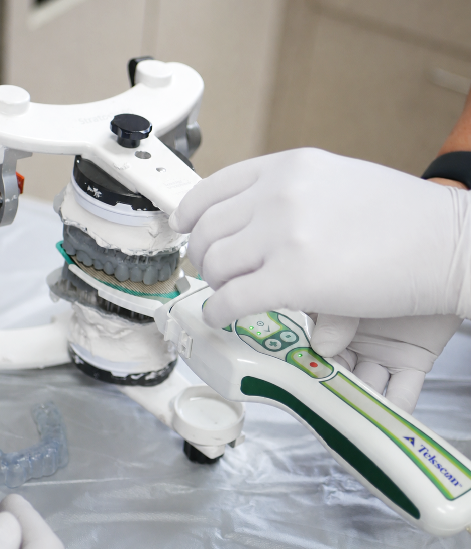
1:30–2:15pm — Lecture: Force management: distribution, concentration, and restorations that withstand the load, demonstrated on digital scans
2:15–3:15pm — Lecture: TMJ disorders: mandibular retrusion, condylar distalization, retrodiscal compression, disc displacement. Assessment and classification: deviation versus deflection and what each indicates, a click versus a pop, and how each finding places the joint in a diagnosis
3:15–3:30pm — Break
3:30–5:00pm — Breakout 3: TMJ assessment and classification in pairs: range of motion, path of opening, and joint sounds, recorded and classified
5:00–6:00pm — Cocktails and hors d'oeuvres
-
8:00–8:30am — Registration and breakfast
8:30–9:45am — Lecture: Frontal and lateral cephalograms: indications, choice of view, head-position protocol, craniocervical angulation, cervical curvature, hyoid, posterior airway. Introduction to CBCT: what it shows, its indications, and basic orientation
9:45–11:00am — Breakout 4: Imaging workshop: frontal and lateral cephalometric interpretation, and basic CBCT reading
11:00–11:15am — Break
11:15am–12:00pm — Lecture: Introduction to appliances: teeth as the stops, the appliance as the replacement stops, the orthotic as the corrective device
12:00–12:45pm — Lecture: From findings to diagnosis and plan. The Treatment-Planning Flowchart
12:45–1:45pm — Lunch
1:45–3:00pm — Breakout 5: Case assessment and treatment planning on 3D-printed models: diagnosis, sequence, maintenance
3:00–3:15pm — Break
3:15–4:00pm — Breakout 6: Presenting comprehensive treatment: role-play using the Breakout 5 plans
4:00–4:45pm — Review of the workshop plans and of cases submitted by attending dentists
4:45–5:00pm — Introduction to Level 3. Evaluations and certificates
Bring your own cases. Attending dentists may submit their own cases for review on Day 2. Instructions are sent after registration.
Hands-on exercises
Mandibular position assessment and bite registration in pairs
Wear facet mapping on 3D-printed models
TMJ assessment and classification in pairs: range of motion, path of opening, and joint sounds
Imaging workshop: frontal and lateral cephalometric interpretation, and basic CBCT reading
Case assessment and treatment planning on 3D-printed models
Presenting comprehensive treatment: patient communication role-play
Your Monday-morning toolkit
Every level ends with a single-page tool you can apply the next working day. It is not a course summary. It is an instrument, built for chairside use by you and your team.
Level 2 - The Treatment-Planning Flowchart.
A one-page decision flowchart that takes you from the completed findings to the treatment you present. It moves through a sequence of branch points, localized problem or systemic, joint involvement present or absent, occlusion stable or unstable, mandibular position correct or not, and resolves to a clear recommendation: single-unit repair, comprehensive restorative treatment, Phase 1 appliance therapy first, or referral. It gives you a repeatable way to decide what to present and the reasoning to defend it.

Bringing a
team member
This course is open to team members. The protocols in this level are taught to be executed by the team, not the dentist alone: records, photography, bite registration, joint assessment, occlusal analysis, and the setup that supports case presentation. When your team member learns them beside you, the workflow is adopted the week you return, rather than taught secondhand and lost. The most common reason continuing education does not change a practice is that the team never trained on it.
The economics of comprehensive treatment
Level 1 links accurate diagnosis to higher-value, correctly indicated treatment. Level 2 turns that diagnosis into a comprehensive plan and protects the result once it is in the mouth. Three drivers follow from the material.
Comprehensive treatment, completed together, serves the patient and the practice. Treating the whole system in a coordinated sequence, rather than one tooth per visit over years, gives the patient a better outcome, a predictable cost, and fewer separate appointments. For the practice, the same patient is more productive: more treatment is diagnosed, presented, and completed, and the schedule fills with planned comprehensive work instead of reactive single visits.
Dentistry that lasts protects the work you have already been paid for. Restorations loaded beyond what they can withstand are redone at your cost, in your chair, against your reputation. Force management converts remakes and repeated adjustments into cases that hold.
Fewer emergencies, fewer surprises. Identifying and addressing functional risk reduces the unscheduled failures that disrupt the schedule and erode patient trust.
From diagnosis
to plan
Level 2 builds on the diagnostic foundation of Level 1 and connects occlusion, force, wear, and joint findings into a comprehensive treatment plan.
New to the Hands-On Series? Start with Level 1 to build the diagnostic foundation first.
Bridge to Level 3
Level 2 introduces the appliance and the mechanism behind it. Level 3 develops appliance therapy in full: design, delivery, and the corrective repositioning of the mandible that Phase 1 treatment depends on.
Have a question before registering?
Not sure which level is right for you, what to expect, or if you should bring a team member? Send us your question and our team will get back to you.

About the instructor
Dr. Agatha Bis has spent 30 years treating complex cases in private practice, with a clinical focus on TMJ disorders, occlusion, restorative dentistry, and the systems that make treatment more predictable. She is the founder of TMJ Whisperer Academy, an educational platform for dentists who want to understand cases more clearly, diagnose with more confidence, and practise with more control.