

Hands-On Dental Course | Level 1
Find the Cause, Not the Symptom
Screening, Examination, and Diagnosis
Diagnose the mechanism behind the breakdown, and change its course.
Level 1 establishes the diagnostic foundation for every case that follows. Most treatment failures are not failures of execution. They are failures of diagnosis: a sound plan built on the wrong starting point. When the diagnosis is wrong, every decision after it moves in the wrong direction.
This level teaches you to gather the right information efficiently, interpret what it means, and reach a diagnosis that reads periodontal, biomechanical, functional, and dentofacial conditions as one integrated picture. You will learn to take a history that explains how the patient arrived at their current state, to separate parafunction-driven breakdown from breakdown caused by caries or restorative failure, and to predict the course of untreated dysfunction: which tooth, which muscle, which symptom presents next. By the close of the two days, you will be able to screen, qualify, examine, and capture the diagnostic records required to plan even complex, full-mouth cases.
Learning objectives
Upon completion, participants will be able to:
Perform a rapid screening and determine when a screening is sufficient and when a comprehensive examination is warranted.
Take a focused history that identifies the events and habits behind the patient's current condition, while filtering out information that does not affect the diagnosis.
Assess periodontal, biomechanical, functional, and dentofacial conditions as an integrated picture rather than as separate findings.
Distinguish parafunction-driven breakdown from breakdown attributable to caries, hygiene, or restorative failure.
Predict the progression of untreated dysfunction: the tooth, muscle, or symptom most likely to present next.
Capture standardized photographic and radiographic records efficiently enough to obtain them on every patient.
Read a panoramic radiograph for functional information, including condylar form, vertical symmetry of the condyle–ramus unit, antegonial morphology, and the dentoalveolar signs of chronic overload, and determine when those findings warrant advanced imaging.
Perform digital scanning and interpret the output within a diagnostic workflow, including the virtual articulator.
Assemble history, examination, imaging, and records into a single defensible diagnosis for complex cases.
Communicate findings and reasonable expectations to patients in a way that supports treatment acceptance.
Course details
Location: Smiles by Bis
710 Dorval Dr #220, Oakville, ON L6K 3V7
Date: January 29 - 30, 2027
CE Credits: 14
Dentist tuition: $1,995 CAD
Hygienist or assistant: $980
Format: Two-day, in-person course
Course content
-
The patient's account, read correctly, explains why they are in your chair. This module teaches you to elicit the information that matters: the sequence of events, the habits, the prior treatment. You will learn which details to probe and which to set aside. The objective is not a longer conversation. It is a more targeted one.
-
Many patients expect outcomes the biology will not support. A mutilated dentition with decades of neglect will not be resolved by a few veneers. This module addresses how to identify what a patient believes is achievable, how their perceived budget shapes that belief, and how to communicate reasonable expectations in a way that moves them toward the appropriate treatment rather than the one they arrived asking for.
-
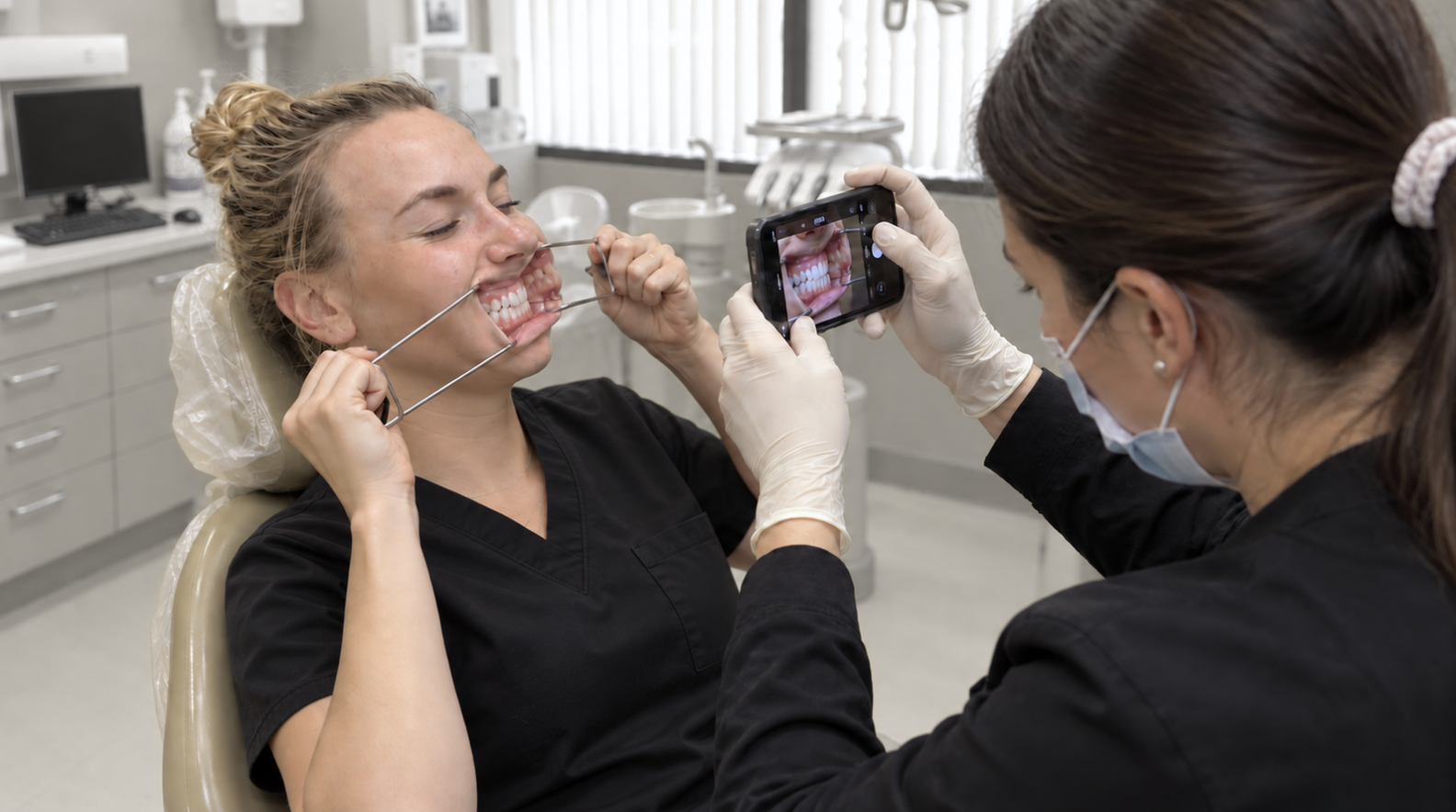
Imaging narrows the diagnosis and the treatment options. Photography drives case acceptance. Both fail in practice when they are slow, hard to delegate, or inconsistent. This module teaches a standardized protocol that your team can perform on every patient, and how to use those records efficiently in a treatment presentation that makes the value of the plan evident to the patient.
-
The panoramic radiograph is taken on nearly every new patient and read for caries, periapical lesions, third molars, and gross pathology. It is then set aside. The same image records the loading history of the masticatory system, and that information is rarely extracted.
This module teaches a second reading of the film. Four findings are covered in detail: condylar form and cortical integrity, vertical symmetry of the condyle–ramus unit, antegonial morphology, and the load signature written into the teeth and alveolar bone. Each is tied to the mechanism that produced it and to the clinical decision it changes.
The module also defines the limits of the image. A panoramic radiograph cannot show disc position and cannot establish condylar position or 3-dimensional shape in the fossa. It raises suspicion and directs further imaging. It does not exclude joint disease. Participants learn which findings warrant CBCT, which warrant MRI, and which warrant neither.
-
Periodontal, biomechanical, functional, and dentofacial findings are usually assessed in isolation. They should be read together. Not all bone loss follows poor hygiene. Not all fractured teeth follow caries or hard food. This module teaches you to identify the parafunction behind the presenting signs and to predict the trajectory of the condition: which tooth, which muscle, which symptom is next if the cause is left uncorrected. Prediction, shown to the patient before it occurs, is what converts a skeptical patient into one who accepts comprehensive treatment.
-
Time spent gathering information on a patient who declines basic treatment is time lost. This module teaches a staged approach: a rapid screening to qualify the patient, a comprehensive examination when the case warrants it, and the diagnostic records that support the presentation and closure of complex treatment. Knowing which step a given patient requires is as important as knowing how to perform it.
-
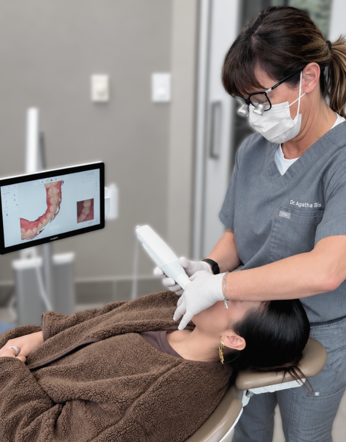
Digital technology runs through every level of the certification. BIS Institute performs all laboratory work onsite, so participants observe the full workflow directly: digital scanning, design, 3D printing, CAD/CAM and milling of single and multiple units, and occlusal analysis with TScan. The program pairs current digital tools with conventional articulators and mounted models, so the material is taught across all four learning modalities: visual, auditory, didactic, and kinesthetic.
-
A treatment plan is only as sound as the diagnosis beneath it. This module brings together history, examination, imaging, and records into one defensible diagnosis. That diagnosis is the deliverable of Level 1 and the required starting point for the treatment planning taught in Level 2.
Hands-on practical exercises
Rapid screening
Comprehensive examination: technique and timing
Critical analysis workshop
Range of motion evaluation
Dental photography: a fast, repeatable protocol
Panoramic interpretation workshop: the four functional findings, read across a case series
Digital scanning: focus on efficiency and tips on how to capture difficult areas
Digital technology introduction: 3D printing, appliance design, CAD/CAM, and occlusal analysis (TScan)
Patient communication role-play
Your Monday-morning toolkit
Every level ends with a single-page tool you can apply the next working day. It is not a course summary. It is an instrument, built for chairside use by you and your team.
Level 1 - The Function-Driven Screening Protocol.
A one-page screening instrument that qualifies every new patient in minutes: the history questions that matter, the TMJ and range-of-motion screen, the parafunction signs to record, the four-point panoramic read, and the threshold that signals when to convert the patient to a comprehensive examination. It standardizes the front end of the diagnostic process so it runs on every patient without adding chair time.
Bringing a team member
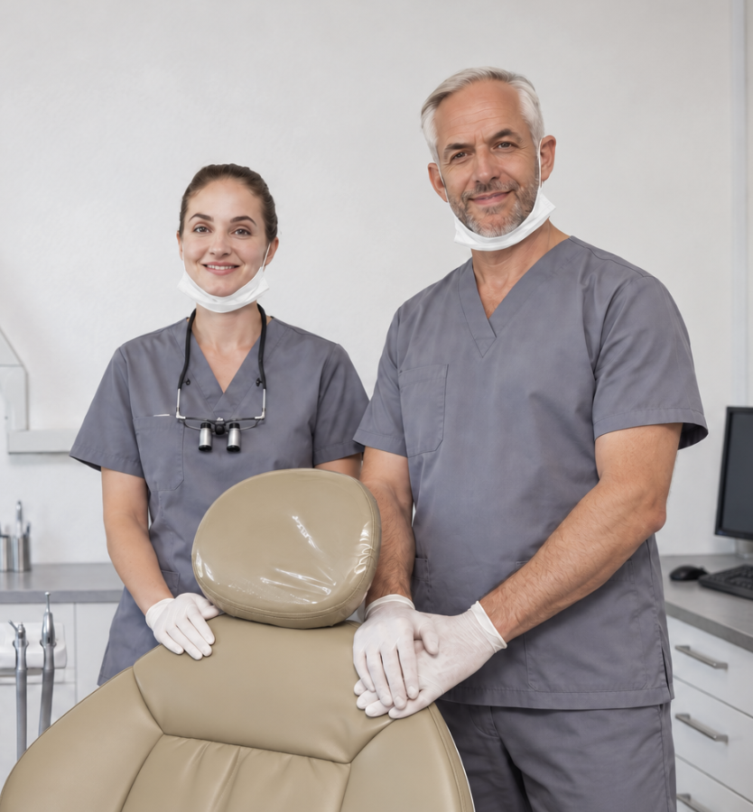
This course is open to all team members.
The protocols in this level are taught to be executed by the team, not the dentist alone: records, photography, charting of clinical findings of parafunction, digital scanning, occlusal analysis, and the setup that supports case presentation.
When your team member learns them beside you, the workflow is adopted the day you return, rather than taught secondhand and lost. The most common reason continuing education does not change a practice is that the team never trained on it.
The economics of accurate diagnosis
Reactive, single-tooth dentistry treats the symptom and returns the patient to the same trajectory. Function-driven diagnosis identifies the cause and the true scope of the case, which is usually comprehensive rather than single-unit treatment. Three consequences follow directly.
Higher-value treatment, correctly indicated. A diagnosed full-arch, occlusal, or orthotic-driven case is a multiple of the single restorations it replaces. The value is not manufactured. It is treatment the patient already needed and that reactive care would have missed. Run the arithmetic against your own fee schedule: one comprehensive case set against the string of single visits it displaces.
Higher case acceptance. Patients accept treatment they understand. When you show a patient the mechanism behind their breakdown and predict what will fail next, acceptance stops depending on persuasion. Each prediction later confirmed also builds the referral base.
Less wasted chair time. Screening qualifies the patient before the comprehensive exam. Diagnostic time goes to the patients who will proceed, not to those who decline basic care.
Ready to take the next step?
Start with Level 1 to build the diagnostic foundation you’ll use throughout the Hands-On series.
Bridge to Level 2
Level 1 establishes the diagnosis. Level 2 builds the treatment plan, working from more complex presentations and introducing the functional and temporomandibular considerations that determine how those cases should be sequenced and treated.
Have a question before registering?
Not sure which level is right for you, what to expect, or whether you should bring a team member? Send us your question and our team will get back to you.

About the instructor
Dr. Agatha Bis has spent nearly 30 years treating complex cases in private practice with a clinical focus on TMJ disorders, occlusion, restorative dentistry, and the systems that make treatment more predictable. She is also the founder of TMJ Whisperer Academy, an educational platform built for dentists who want to understand cases more clearly, diagnose with more confidence, and practice with more control.